

“Milia” is also widely used in Turkey as “Milialar.” It describes the tiny, pearl-like cysts that frequently develop on the skin surrounding the eyes, especially the eyelids. Even though these tiny lumps are usually benign, their appearance can cause anxiety. Remember that milialar acne is not the same as other types of acne. In this thorough guide, we shall examine the realm of milialar, also known as milia, in this guide, looking at its kinds, progression, indications, symptoms, precautions, and available treatments. You may be sure you completely understand Milia because the information is based on in-depth scientific studies and professional perspectives on the illness.
Milia or Milialar: what is it?
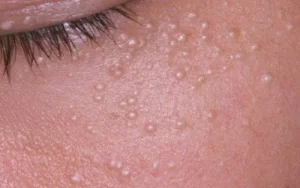
Milialar are tiny, pinhead-sized, dome-shaped lumps that usually range from 1-2 millimetres. On the skin’s surface, they resemble firm, smooth, pearly, whitish-yellow cysts. Milia most frequently appears on the eyelid and in the area beneath the eyes, resembling microscopic pearls buried in the skin. A study claims that milialar happens when keratin, a protein in skin, hair, and nails, gets trapped under the skin’s outer layer. Although they are frequently observed in infants, milialar can also form in adults, usually due to skin injury.
Milia or Milialar types
Primary and secondary milialar are the two categories into which milialar can be divided.
Milialar or Primary Milia
Pediatric milialar is directly produced by keratin being trapped in the skin. Because of their underdeveloped sweat ducts, neonates are more likely to have them. Important traits of primary milia, also known as milialar, consist of:
- Tiny, yellow-to-white cysts.
- Frequently observed on the face, particularly the nose, cheeks, and eye area.
- Usually symptomless.
- In newborns, this usually goes away on its own in a few weeks or months.
Milialar or Secondary Milia
Conversely, secondary milialar develops as a result of skin damage or injury. Adults may experience them following specific skin surgeries or disorders. Important traits of milialar, or secondary milia, include:
- Comparable in appearance to primary milialar, frequently observed in regions affected by surgery or trauma.
- Some symptoms, such as burn pain, may be related to the underlying cause.
- The length of time varies and may last longer based on the cause.
- It is imperative to address the underlying cause, and therapies such as laser therapy, manual extraction, or medication may be contemplated.
What Leads to the Development of Milialar or Milia?
Dead skin cells get stuck beneath the skin’s surface and create microscopic cysts, which is how milialar originates. They can occur anywhere on the body, though they frequently appear on the face, particularly around the eyes and cheeks. Although the fundamental cause of milia or milialar development is unclear, several factors are involved. Among these are:
- Genetics: Milia is a condition that some people are born with and frequently runs in families.
- Sun Exposure: Prolonged sun exposure raises the risk of milia by causing skin damage to the face over time.
- Skin Trauma: During the healing process, milia may occur on the skin due to wounds, burns, abrasions, and blisters.
- Certain Medical Conditions: Eczema and other conditions that produce dry skin and irritation may make you more susceptible.
- Medication: Steroids, for example, have the potential to cause milia as a side effect.
- Thick Creams and Makeup: Using greasy, thick makeup can clog pores and result in cysts.
Newborns are most likely to acquire milia, with up to 50% experiencing temporary milia that usually disappears in a few weeks. It is thought that maternal hormones have a part in this phenomenon. Nonetheless, 2.5% of all adults are affected by persistent milialar illness. Age-related changes in skin cell kinetics and decreased skin flexibility are assumed to cause milia, which affects women more often than males and becomes more common as people age.
Method of Development
Milialar is developed by a particular procedure:
- Skin Renewal: The skin naturally sheds dead cells as part of this process. These cells don’t always shed correctly.
- Trapped Keratin: After being trapped, the cells produce keratin, which builds up.
- Cyst Formation: As a result of this accumulation, microscopic cysts under the skin’s surface form, which causes milia.
Symptoms and Indications of Milialar or Milia
Milialar is easily recognized in most cases because of its distinctive look. They may appear as follows:
- Little white pimples on the eyelids or surrounding the eyes.
- Pearl-like, smooth, dome-shaped lumps beneath the skin.
- Color: whitish-yellow or yellowish-white.
- May show up alone or in groups.
- They don’t hurt and usually don’t itch or irritate.
- Can either vanish on their own or stay unchanged for several weeks or months.
- May occasionally, if burst, release a waxy, cheese-like secretion.
Preventive Actions
Although milialar development may not be completely preventable, the following advice may help lower the likelihood that they will occur:
- Use makeup and moisturizers that are non-comedogenic and oil-free.
- Steer clear of greasy, thick lotions and makeup near your eyes.
- To clear clogged pores, gently cleanse your face and exfoliate it frequently.
- Take care during shaving, employing the right method to prevent skin injuries.
- Limit exposure to the sun without protection and use sunscreen every day.
- Keep your skin hydrated to avoid being overly parched.
- Take off all makeup well before bed, and throw away any leftovers.
- Address any underlying skin diseases, such as eczema.
- If you have milia, also known as Millia, you should avoid chemical peels or intense facials since they might exacerbate the condition.
Options for Milia or Milialar Treatment
Milialar rarely needs to be treated; many cases go away independently in weeks or months. However, there are a few therapeutic choices available if the bumps continue or create discomfort:
- Prescription Retinoid Creams: The milia can be dried out and shed using creams including tretinoin, adapalene, or tazarotene.
- Microdermabrasion: This method gently exfoliates the outer layers of skin and promotes healing using tiny crystals.
- Chemical Peels: To soften and eradicate the lesions, apply a moderate glycolic or salicylic acid solution.
- Electrocautery: Using a local anesthetic, a hyfrecator cauterizing device is used to burn off the milialar.
- Manual Extraction: A dermatologist can use a sterile needle to crack open the cyst and extract its contents.
- Cryotherapy: Using liquid nitrogen to freeze the lumps to remove the lesions.
- Laser ablation: The cysts are destroyed by laser energy.
- Surgical Excision: A dermatologist may choose to surgically remove the milia by making an incision, draining the milia, and occasionally sutures.
In summary
Finally, milialar, or tiny pearl-like cysts that can develop on the skin, are located on the eyelids and surrounding tissues and are usually benign. Adults can also get them, though they are more prevalent in infants, usually due to skin injury. There are several varieties of milialar, and variables such as heredity, sun exposure, trauma to the skin, illnesses, drugs, and the use of thick lotions and makeup can all impact how they grow.
There are several therapy options available if you have milialar, even though many cases go away on their own. It’s critical to follow good skincare procedures, stay away from heavy makeup, avoid wearing makeup altogether, and shield your skin from too much sun exposure if you want to prevent milia.
Thanks to this extensive guide, you now understand milia thoroughly, including its kinds, development, signs and symptoms, prevention measures, and available treatments. See a dermatologist to get expert advice if you’re worried about your skin or nails.


